
Mitochondria: Morphology, structure and Function
Mitochondria (Gr: mito = thread; chondrion =
granule) (Singular: mitochondrion) are most essential cell-organelles found
distributed in the cytoplasm of plant and animal cells and protozoa.
However, they are not found in bacteria and red blood cells of multicellular animals.
Mitochondria are characterized by a number of morphological, biochemical and functional
properties like, size,shape, special staining properties, the specific
structural organization, lipoprotein composition, and presence of certain
specific enzymes and coenzymes.
From the physiological viewpoint, mitochondria are known
as "Biochemical machines “because they recover the energy contained in the
food stuffs through Kerb's Cycle and the respiratory chain. The energy is
produced in the form of ATP (adenosine-triphosphate) by the process phosphorylation. ATP possesses the high
energy phosphate bond. Most commonly mitochondria are also known as Power-House
of the cell because they produce the energy necessary for many cellular
functions.
- HISTORY
Mitochondria were first observed by Kollicker in
1850. Flemming (1892) named them 'fila', Later on Altmann (1894) described them
as bioblasts. Benda (1897) named them asmitochondria. Michaelis (1900) stained
them supravitally with janus green. Regard (1908) established their chemical
nature. Kingsbury (1912) suggested that mitochondria were the sites of cellularoxidation.
Lewis and
Lewis (1914) observed their sensitivity to metabolic conditions. Mever (1918)
described their transformation into various other type of cells. Keilin (1923)
demonstrated the effects of cyanide and carbon monoxide on respiration. Hogeboom,
et al. in (1948) finally confirmed that mitochondria were indeed the sites of
cellular respiration. Very recently it has confirmed that mitochondria contain
a specific type of DNA different from that of nuclear DNA.
They perform the process of protein synthesis in
them and also take part in the genetical phenomenon of "Plasma
inheritance". This plasma-inheritance where the characters are transmitted
through the mitochondria of female, is known as mitochondrial inheritance.
- MORPHOLOGY:
shape:The shape of mitochondria varies according to the
type of cell, In general, they are sausage shaped or filamentous and rod-shaped organelles that can be considered the power generators of the cell,
Depending
upon the physiological conditions they may be club-shaped, tennis
recket-shaped, rod-shaped or vescular in shape. Such shapes are only for a
short period and after 48 hours these changes cease and the mitochondria regain
their original form.
SIZE:
The size of mitochondria is also variable and
chiefly depends upon the functional stage of the cell. The length of
mitochondria varies from 1.5 u to 7 u and the width is relatively constant being
0.5 u. Very thin mitochondria are about 0.2 u and thick about 2 u. In fixed
preparations their size and shape depend upon osmotic pressure and pH of the
fixative. In acidic pH they tend to fragment and to become vescicular. Mitochondria of rat-liver are 3.3. u in length; of mammalian exocrine pancrease
about 10 u in length; and of amphibian cocytes about 20 to 40 u in length.
NUMBER:
Like shape
and size, number of mitochondria also varies according to the type and functional
activities of the cell. Generally, a cell has 200 to 300 mitochondria but their
number may reach from a few to 1000 or more. Some algal cells have been
reported to have only one mitochondria. A normal liver cell possesses about
1000 to 16,000 mitochondria; eggs of sea urchin 14,000 to 150,000; ovocytes
300,000; and Chaos about 500,000 mitochondria. The animal cells posses greater
number of mitochondria than green plant cells. Mitochondria are rod-shaped organelles that can be considered
the power generators of the cell.
- LOCATION:
- ORIENTATION:
Mitochondria possess a more of less definite orientation in some
cells. For example, in cylindrical cells, their orientation is in the
basal-apical direction, parallel to the main axis. In leukocytes, their
arrangement is redical with respect to the centrioles. The orientations of mitochondria
depend upon the direction of the diffusion currents within cells (Pollister,
1941) and are also related to the sub-microscopic organisation of the
cytoplasmic matrix and vacuolar system.
Structure of Mitochondria:
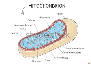
Structure of Mitochondria:
A
mitochondrion contains two membranes and p,g g 34 structure of a mitochondrion,
two chambers, outer and inner. The A mitochondrion partly cut open to show two
membranes form the envelope of the mitointernal and external structure,
chondrion. Each of them is 60-75A in thickness.
Outer Membrane:
The
membrane is smooth. It is permeable to a number of metabolites. It is due to
presence of protein channels called porins or minute pores. A few enzymes
connected with lipid synthesis are located in the membrane. It is poorer in
proteins as compared to inner membrane.
Inner Membrane:
It
is permeable to only some metabolites. It is rich in double phospholipid
called cardiolipin (having four fatty acids) which makes the membrane
impermeable to ions. Protein content is also high, being 70—75% of total
components. The inner membrane is in-folded variously to form involutions
called cristae. They are meant for increasing the physiologically active area
of the inner membrane.
The
cristae are generally arranged like baffles, at right angles to the
longitudinal axis of the mitochondrion. They are tubular (most plant cells) or
plate like (most animal cells) or vesicle-like (e.g., Euglena). A crista
encloses a space that is continuation of the outer chamber. The density of
cristae indicates the intensity of respiration.
A
channel occurs between roter and stator for passage of protons (H+). Stator is
connected to head region by an arm. Enzymes of electron transport are located
in the inner membrane in contact with elementary particles.
At
places, outer and inner mitochondrial membranes come in contact. They are
called adhesion sites. Adhesion sites are special permeation regions of the
mitochondrion for transfer of materials from outside to inside and vice versa.
Outer Chamber (Peri-mitochondrial Space):
The
chamber is the space that lies between the outer and inner membrane of the
mitochondrial envelope. Usually, it is 60-100 A wide. It extends into the spaces
of the cristae.The chamber contains a fluid having a few enzymes.
Inner Chamber:
It
forms the core of the mitochondrion. The inner chamber contains a semi-fluid
matrix. The matrix has protein particles, ribosomes, RNA, DNA (mitochondrial or
mDNA), enzymes of Krebs or TCA cycle (except succinate dehydrogense which is
membrane based), amino acid synthesis and fatty acid metabolism, crystals of
calcium phosphate and manganese.
Mitochondrial
ribosomes are 55 S to 70 S in nature. They thus resemble the ribosomes of
prokaryotes. DNA is naked. It is commonly circular but can be linear. DNA makes
the mitochondrion semi-autonomous.
Functions of Mitochondria:
1.
Mitochondria are miniature biochemical factories where food stuffs or
respiratory substrates are completely oxidized to carbon dioxide and water. The
energy liberated in the process is initially stored in the form of reduced
coenzymes and reduced prosthetic groups.
The
latter soon undergo oxidation and form energy rich ATR ATP comes out of
mitochondria and helps perform various energy requiring processes of the cell
like muscle contraction, nerve impulse conduction, biosynthesis, membrane
transport, cell division, movement, etc. Because of the formation of ATP, the
mitochondria are called power houses of the cell.
2.
Mitochondria provide important intermediates for the synthesis of several
bio-chemicals like chlorophyll, cytochromes, pyrimidine’s, steroids, alkaloids,
etc.
3.
The matrix or inner chamber of the mitochondria has enzymes for the synthesis
of fatty acids. Enzymes required for the elongation of fatty acids have been
reported in the outer mitochondrial chamber.
4. Synthesis of many amino acids occurs in
the mitochondria. The first formed amino acids are glutamic acid and aspartic
acid. They are synthesized from a-ketoglutaric acid and oxaloacetic acid
respectively. Other amino acids are produced by transformation and transamination
or transfer of amino group (—NH2) from glutamic
acid and aspartic acid.
5.
Mitochondria may store and release Calcium when required.
6.
An organism generally receives mitochondria from its mother (maternal
inheritance).
REFERENCE
1. Logan DC (June 2010). "Mitochondrial fusion,
division and positioning in plants". Biochemical Society
Transactions. 38 (3):
789–95. doi:10.1042/bst0380789. PMID 20491666.
2.
das Neves
RP, Jones NS, Andreu L, Gupta R, Enver T, Iborra FJ (December 2010). Weissman
JS (ed.). "Connecting variability in global
transcription rate to mitochondrial variability". PLoS
Biology. 8 (12):
e1000560. doi:10.1371/journal.pbio.1000560. PMC 3001896. PMID 21179497.
3.
Johnston
IG, Gaal B, Neves RP, Enver T, Iborra FJ, Jones NS (2012). Haugh JM
(ed.). "Mitochondrial variability as a
source of extrinsic cellular noise". PLoS Computational
Biology. 8 (3):
e1002416. arXiv:1107.4499. Bibcode:2012PLSCB...8E2416J. doi:10.1371/journal.pcbi.1002416. PMC 3297557. PMID 22412363.
4.
Rappaport
L, Oliviero P, Samuel JL (1998). "Cytoskeleton and mitochondrial
morphology and function". Mol. Cell. Biochem. 184: 101–105. doi:10.1023/A:1006843113166.
5.
Hoitzing
H, Johnston IG, Jones NS (June 2015). "What is the function of
mitochondrial networks? A theoretical assessment of hypotheses and proposal for
future research". BioEssays. 37 (6): 687–700. doi:10.1002/bies.201400188. PMC 4672710. PMID 25847815.
6.
Soltys BJ,
Gupta RS (1992). "Interrelationships of endoplasmic reticulum,
mitochondria, intermediate filaments, and microtubules--a quadruple
fluorescence labeling study". Biochemistry and Cell Biology. 70 (10–11): 1174–1186. doi:10.1139/o92-163. PMID 1363623.
7.
Tang HL,
Lung HL, Wu KC, Le AH, Tang HM, Fung MC (February 2008). "Vimentin
supports mitochondrial morphology and organization". The Biochemical
Journal. 410 (1):
141–146. doi:10.1042/BJ20071072. PMID 17983357.
8.
Rich PR
(December 2003). "The molecular machinery of Keilin's respiratory
chain". Biochemical Society Transactions. 31 (Pt 6): 1095–1105. doi:10.1042/BST0311095. PMID 14641005.